
Sample Preparation
Cressington 108 Sputter Coater
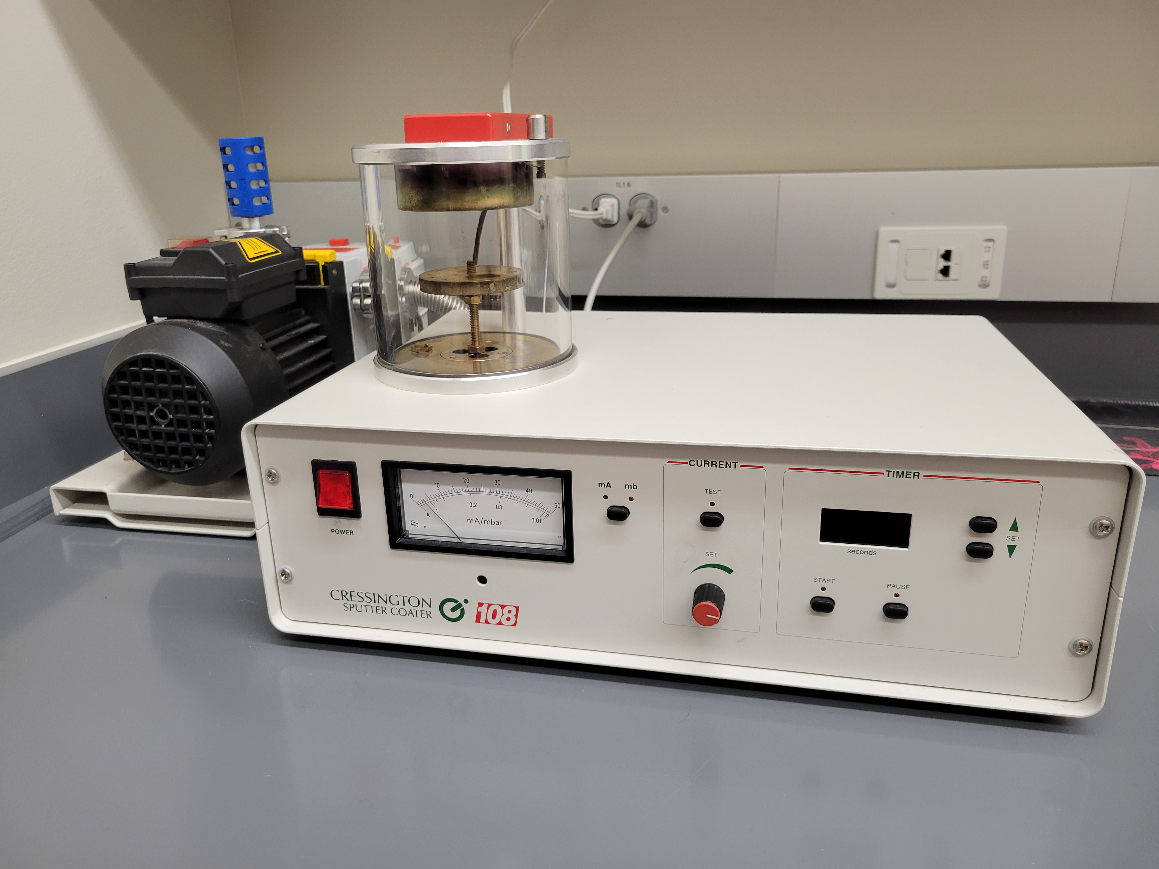
Electron microscopy can pose some complications for biological samples. Due to most organic material being made up of elements with a low atomic number, performing scanning electron microscopy on unprepared organic tissue could result in low signal and a bad image altogether. To compensate for this, sputter coaters are designed to deposit a fine film of gold particles on top of the sample studs. This thin layer of gold (sometimes platinum for higher magnification SEM) helps to distribute electron charge to improve contrast, as well as increase signal from secondary electrons.
The Cressington 108 Sputter Coater in the prep room is capable of coating 12 small sample studs in one cycle. Time spent sputtering, pressure, and sputter current can all be adjusted to accommodate a variety of sample surfaces.
Quorum E3100 Critical Point Drier
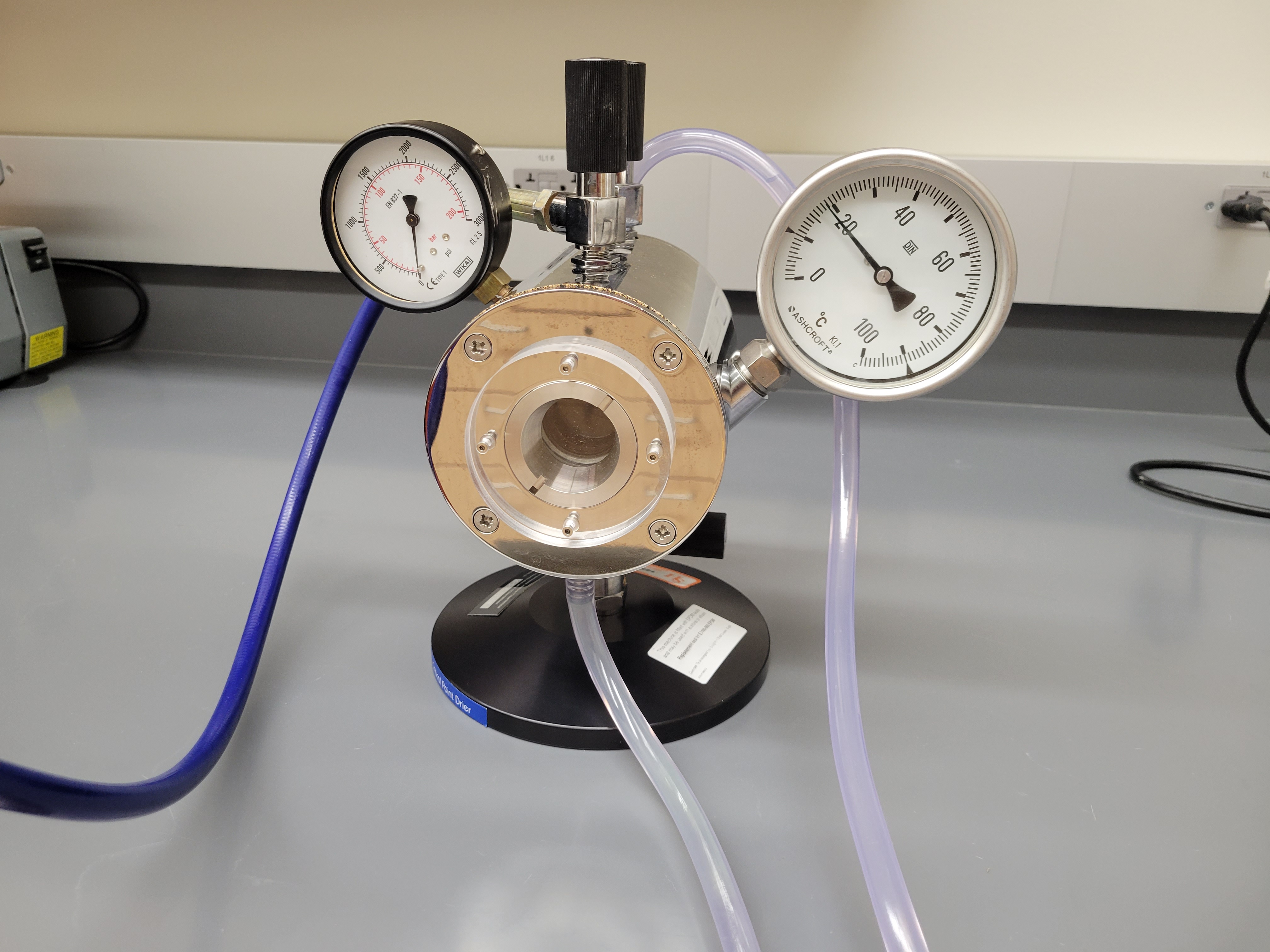
Maximum # of sample baskets: 25 baskets
Lowest Temp. with ice bath: 9°C
Safety Burst: 1825 psi
For particularly small samples, a thin layer of gold won't be enough to produce good images with SEM. Many fine structures may experience warping when allowed to dry in the air, surface tension can cause tissues to shrivel up this way. Additionally, samples that are not completely dry may experience spot heating when exposed to the electron beam. Depending on the size of the sample and how dry it is, sometimes dramatic warping and sometimes burning of the sample can occur due to the intensity of the electron beam.
To account for this, the critical point drier is a completely manual apparatus that uses the critical point of carbon dioxide to exchange wet intermediate fluid in the samples for carbon. This enables small samples to completely dry without any artifacts during imaging from either air drying or high electron beam intensity.
Leica UC7 Ultramicrotome
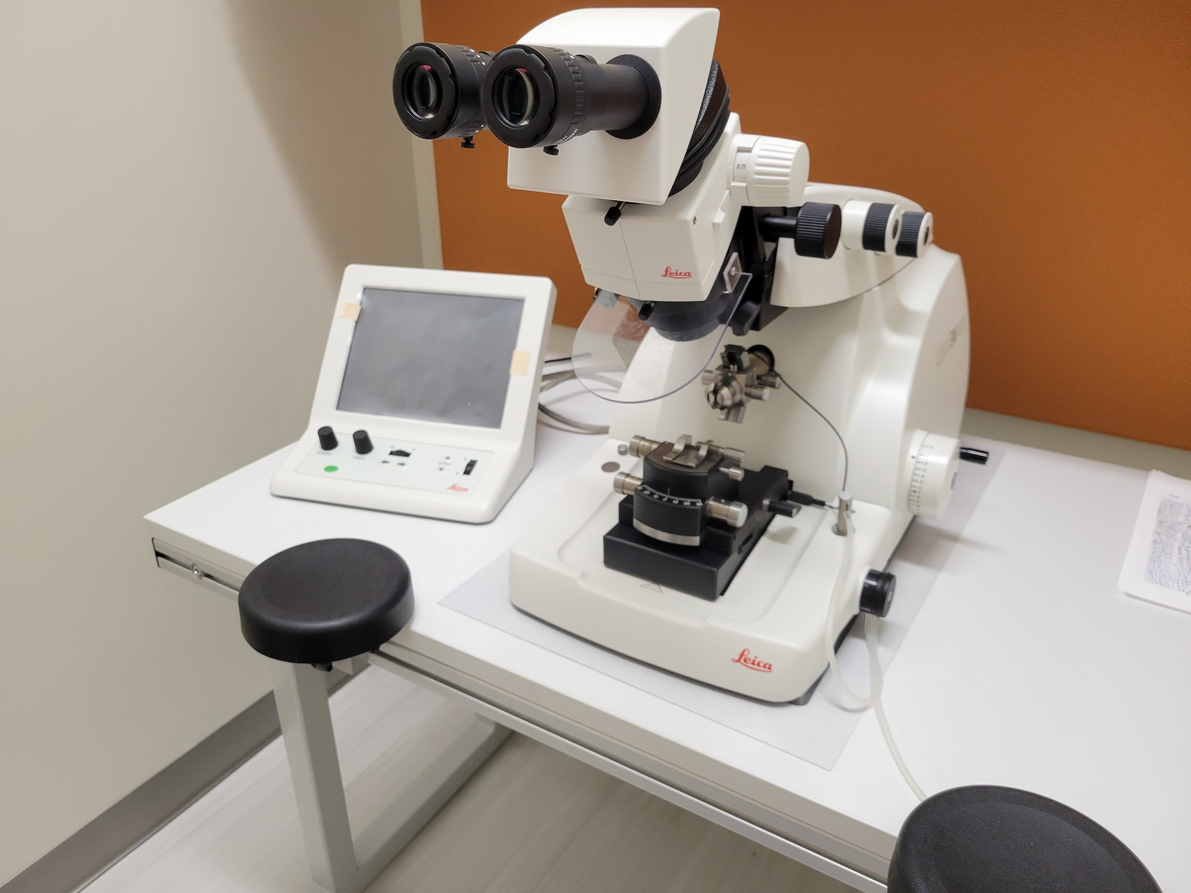
Section Range: 1µm - 50nm
Thin sectioning is necessary for both transmission electron microscopy and confocal microscopy techniques. Ultramicrotomy can achieve sections as thin as 50nm by slowly shaving off pieces of a resin block using a glass knife. These thin sections are much easier to work with than large samples which may be too thick for TEM, where the electron beam may not provide sufficient contrast. SEM and light microscopy can also benefit from sectioning. When studying a particular tissue or even organelle within a cell, it may be useful to cut several sections to assure that the best possible image is obtained.