X-Ray Analysis
X-Ray Analysis
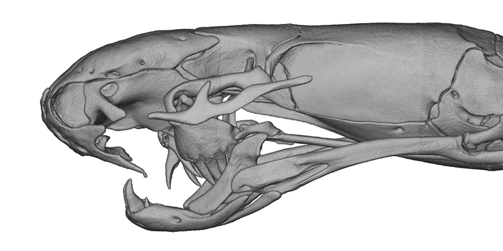
Boasting a higher resolution than standard light microscopy, X-ray imaging has become increasingly common with the advent of high contrast CT (Computed Tomography) methods such as dice-CT. These technologies make three-dimensional visualization possible by imaging an entire volume at once, which is subsequently sectioned digitally to reproduce a reconstructed skeleton, circulatory system, dense nerve fibers, and more.
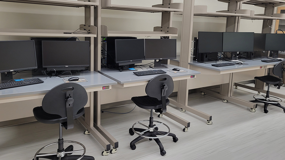
Many undergraduate students take on research projects utilizing the CT analysis lab, sometimes leading to peer-reviewed article publications. As open-access volumes of data become more widely available online, the possibilities exponentially increase each year for independent discovery. Graduate students may take advantage of a visual methods course offered once annually to gain competency in modern High-Resolution CT reconstruction software.
X-Ray / CT Analysis Careers
While specialization is required for X-ray imaging in healthcare, core theory and image interpretation can be learned with preserved animal specimens here at the MCF. Additionally, the usage of CT reconstruction is prevalent in medical research and computer science integrations. As equipment and methods continue to develop, a foundation in this field is increasingly marketable.