Light Microscopy
Fluorescence Microscopy
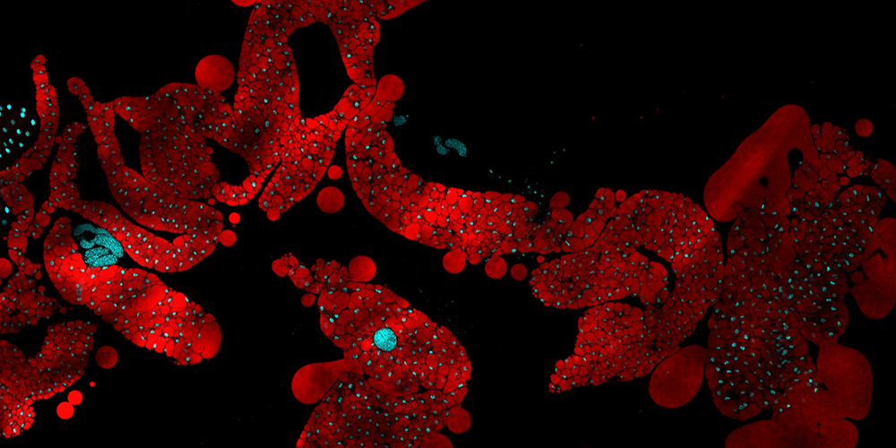
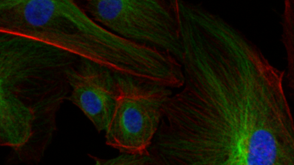
Use advanced staining to visualize molecular systems. Several ECHO Revolve microscopes allow for both upright and inverted imaging with ease - aided by a standard tablet display. Three available LED channels target common excitation wavelengths for undergraduates to become accustomed to fluorescent imaging.
Take advantage of the FLUOVIEW FV3000 confocal laser scanning microscope by Olympus to see three-dimensional volumes of slide-mounted tissue or culture plates. This high-powered scope system uses scanning to limit out of focus light and results in a detailed image with sharp edges to fluorescent elements.
Different stains (called fluorophores) are available to target specific cellular components and provide discrimination to the researcher. The lasers of our FV 3000 system are detailed below by wavelength so users can prepare their methods accordingly.
Available Lasers:
- 405
- 445
- 488
- 514
- 561
- 640
Visible Light Microscopy
Use our high-powered KEYENCE VHX microscopes to accomplish visible light data collection like never before. The user-friendly interfaces of these systems take advantage of relevant post-processing effects; all available right at the lab bench to save time for the user.
Two VHX series scopes are available for undergraduate and graduate students alike to acquire data at their convenience. These workhorse microscopes are commonly responsible for several research publications each academic year.
VHX Software Functions:
- Depth Composition: Create a high-resolution composite of quickly captured image stacks to remove blurry elements from the field of view.
- Image Stitching: Capture large, pixel-dense images by adding together many fields of view quickly.
- Post-Processing: HDR and Glare removal (among other effects) can improve final image quality for difficult samples.
- Area and Count Statistics: Perform measurements immediately upon capturing an image, including density and circularity analysis.
Light Microscopy Careers
The basis of most biological imaging, light microscopy continues to provide a strong foundation for students of biology. Advances in fluorescence, such as Super-resolution methods, build on the basic theory and practice which students can learn through both undergraduate research and coursework-integrated experiences.
Premed students will benefit greatly from histology, basic sample preparation, and visible light methodology. Students looking for a career in biotechnology or advanced clinical research should look to gain experience during their time at SHSU with a variety of light microscopy methods within the MCF.