SHSU Microscopy Center
The Microscopy Center at SHSU is devoted to making all sorts of visual data available to faculty and students alike. High powered electron microscopes, laser scanning and wide field fluorescence scopes, digital microscopes, and more are all used for impacting research at Sam in a variety of fields. Though labs are found on the first and second floor of the Life Sciences Building, these machines see use from departments across the College of Science and Engineering Technology.
An advanced microscopy course (BIOL 5394) is available to graduate students interested in learning some of the equipment available, offered by Dr. Justin Williams and Daniel Doucet during the fall. Scanning Electron Microscopy, an emphasis of the course, sees application beyond biology - often used by industry for quality assurance and research/development.
Contact: For equipment details, plans to visit, or other questions, please email our Microscopy Lab Coordinator, Daniel Doucet: doucet@shsu.edu
Your Resources
HITACHI SU3500 Scanning Electron Microscope
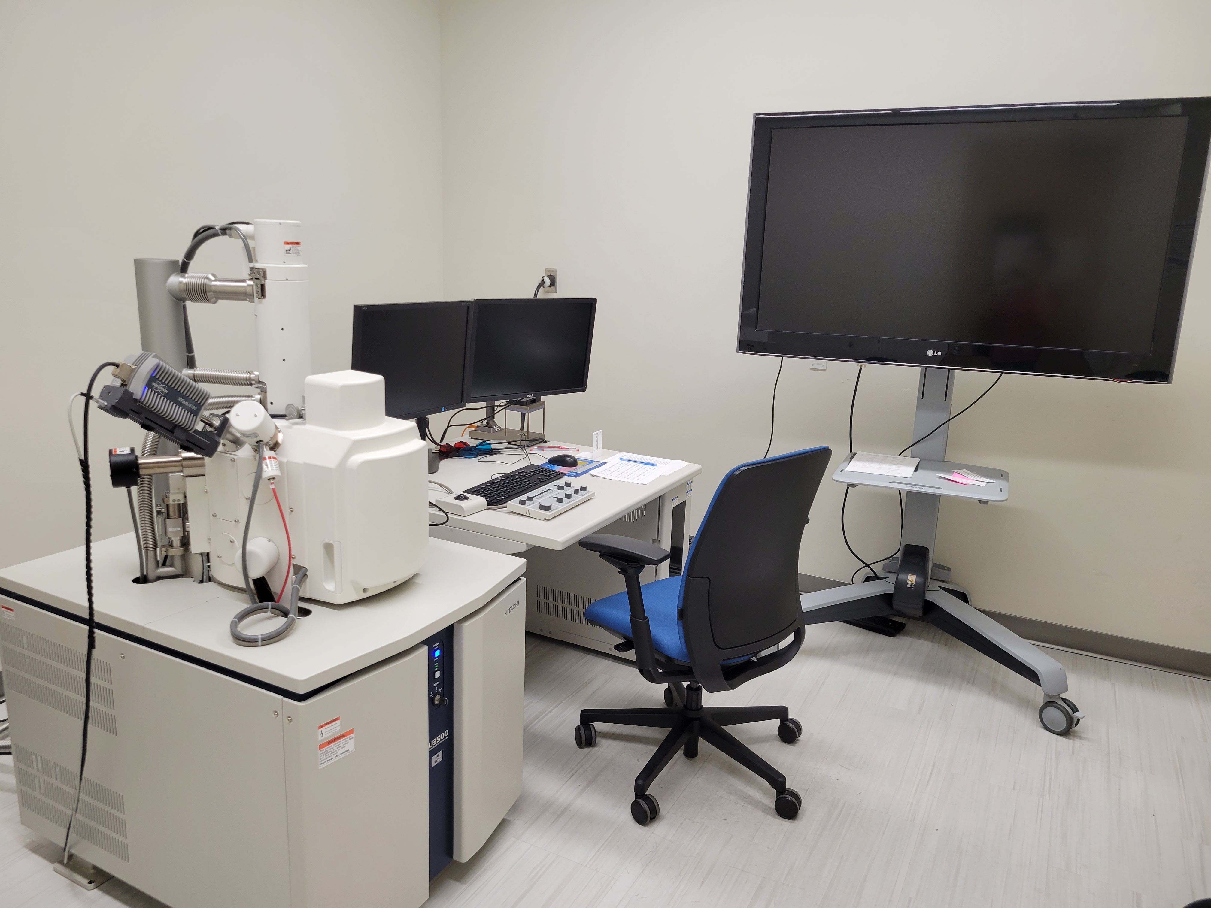
Filament: Tungsten hairpin, >30 µm source diameter (~100 hr lifetime)
Detectors: Secondary Electron (SE), Backscatter Electron (BSE), Bruker QUANTAX EDS (EDS)
Resolving Power: 50 nm @ 1 kV, 4 nm @ 30 kV
With a variety of spot intensity and accelerating voltage (1 to 30 kV), the SEM allows for careful, yet high-resolution imaging of many different sample surfaces. Proper preparation of biological samples allows even delicate samples to be imaged with the electron beam at lower voltage and vacuum. BSE and EDS detectors allow for qualitative and quantitative analysis of element composition. Our SEM has a particularly simple user interface, meaning even inexperienced students to feel comfortable taking publication-quality images within a few hours.
HITACHI H-7500 Transmission Electron Microscope
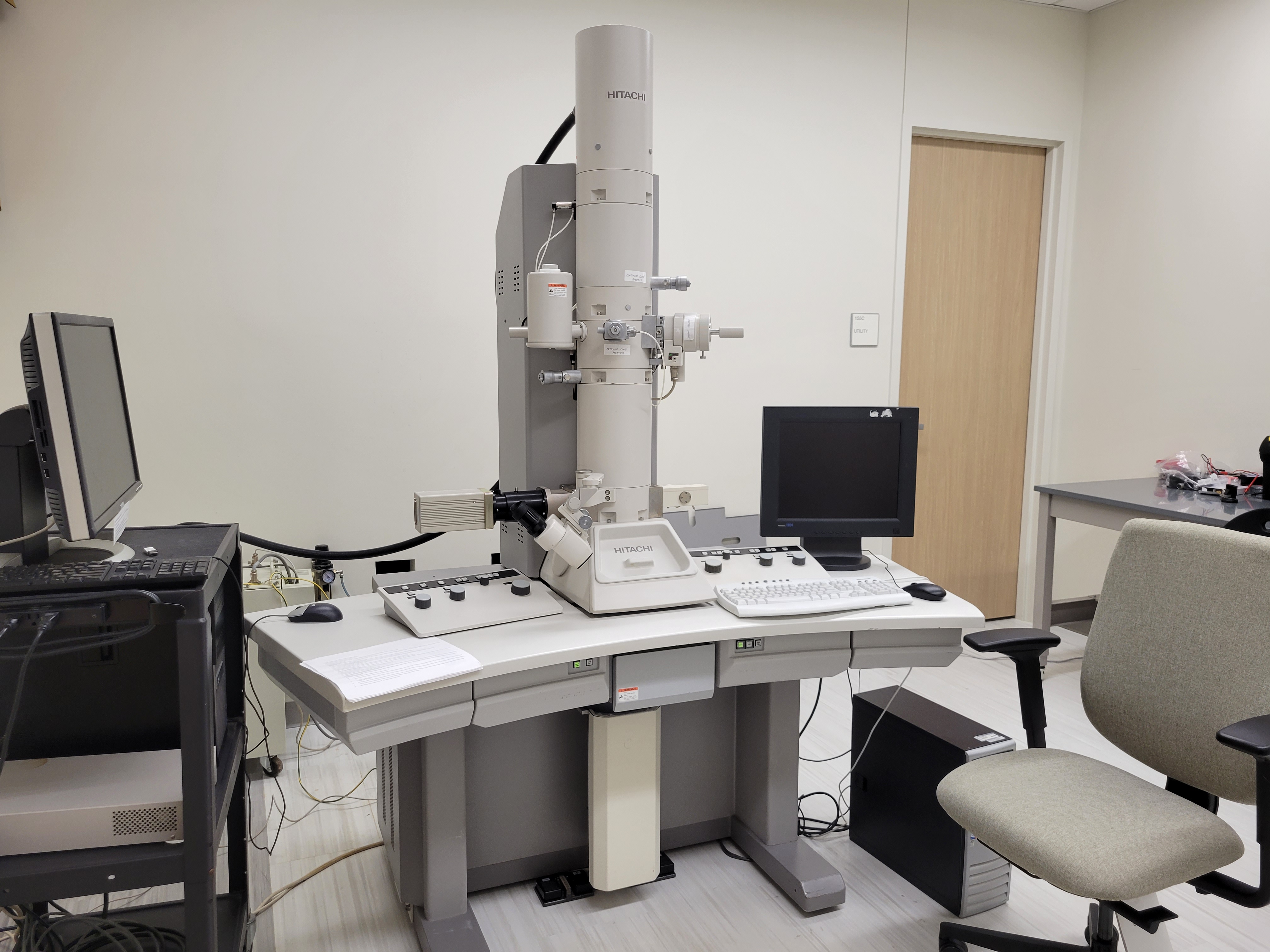
Filament: Tungsten hairpin (~300 hr lifetime)
Resolving Power: 0.23 nm
Accelerating Voltage: 100 kV (20-120kV)
Capable of a finer resolution than most scanning electron microscopes, the TEM relies on instead shooting electrons through the sample to visualize morphological information. This scope typically sees a particular niche compared to the SEM, which can see application in a number of different disciplines and study areas. Chemical and materials science particularly benefit from the high resolving power of this scope.
Biological samples for the TEM are typically cut especially thin (less than 1 µm) to allow for distinct contrast to appear. Sample preparation is often the most time-consuming part of TEM imaging. It is necessary for organic tissues to be fixed to not only stabilize the cells and any organelles within, but to provide enough contrast for the electron beam to distinguish between separate structures.
FLUOVIEW FV3000 Confocal Microscope
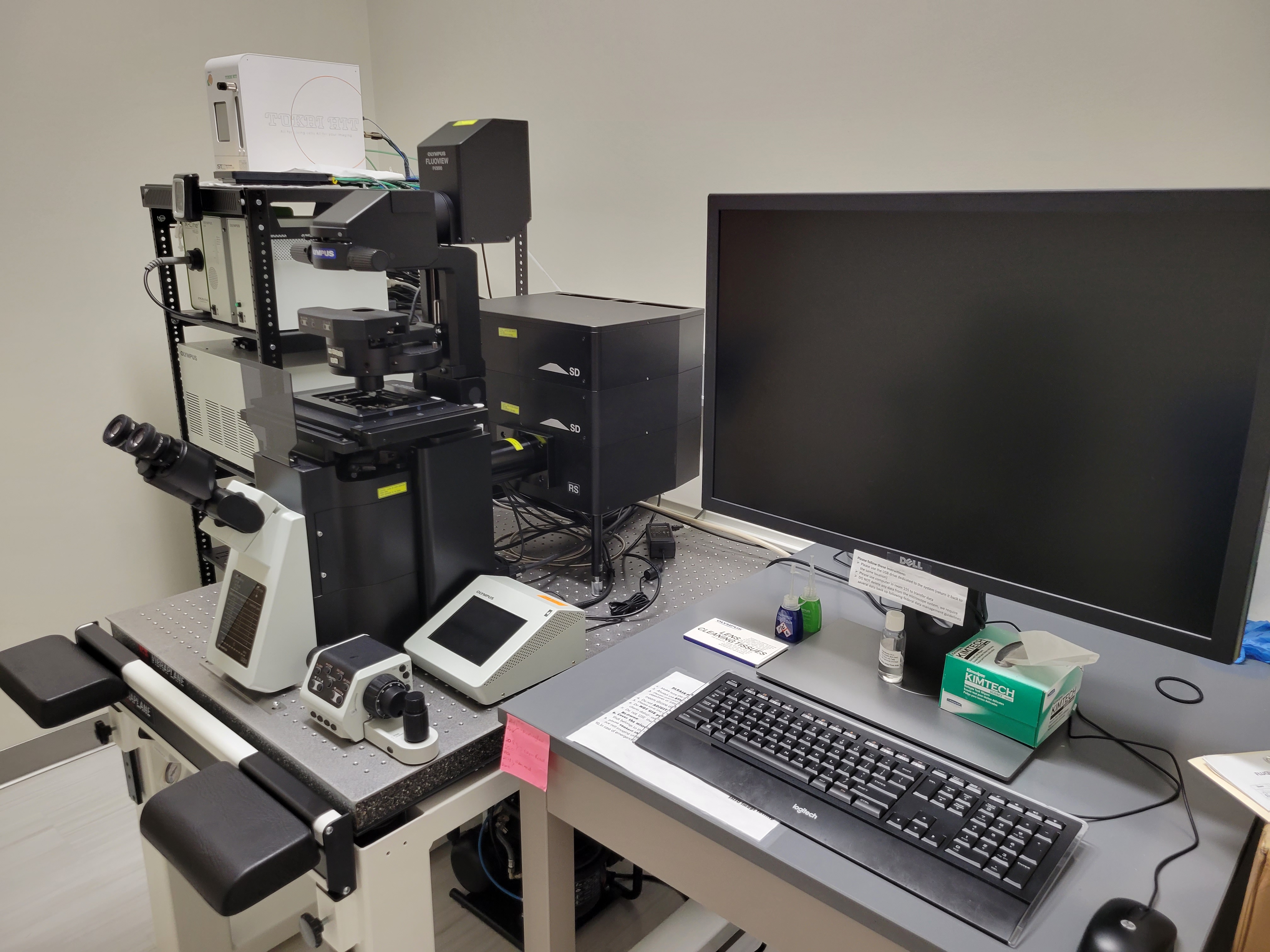
Lasers (avg. wavelength underlined): OBIS405-50LX, OBIS445-75LX, OBIS488-20LS, OBIS514-40LX, OBIS561-20LS, OBIS594-20LS, OBIS640-40LX
Objective Lenses (magnification underlined): UPLSAPO10X2, UPLFLN10X2, UPLFLN10X2PH, UPLFLN20X, UPLFLN20XPH, UPLFLN40X, UPLFLN20XPH, CPLFLN10XPH, UCPFLN20X, LUCPLFLN20XPH, LUCPFLN40X, LUCPFLN40XPH, LUCPFLN60X, LUCPFLN60XPH
A Confocal Laser Scanning microscope works by directing high-energy lasers to singular points on the sample, then rastering the laser across the sample surface. Extremely high-quality images are produced by measuring the emitted or reflected light returned from the scan. The FLUOVIEW FV3000 is also capable of filming videos due to its potential for high scan speeds. Various channels for each laser and emitted light result in several images which can either be combined into one picture or viewed on separate panels to visualize exactly where each fluorophore is present.
KEYENCE VHX-5000 / KEYENCE VHX-600
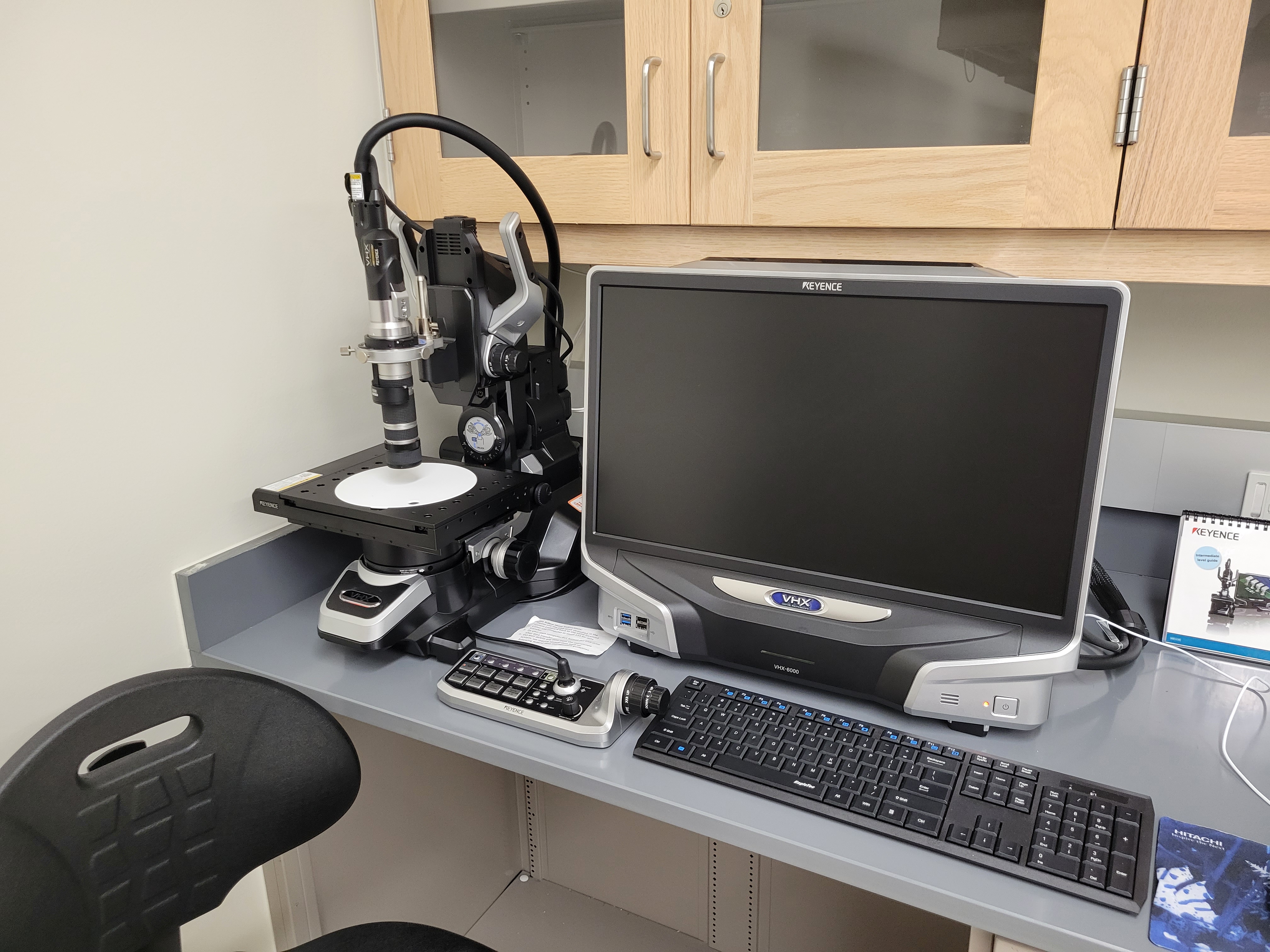
Magnification: Standard Lens, 20-200x / Wide Lens, 5-50x
Image Quality: 1612 (H) x 1212 (V) per capture / max size of 20000 pixels (H) x 20000 pixels (V) with stitching
KEYENCE digital microscopes allow for several approaches to imaging. The powerful VHX-5000 has a 1.95 million pixel CMOS image sensor, providing extremely high resolution images - made even more useful with its image stitching function, where several pictures are taken of an area (using the computer-controlled moving stage) and subsequently combined in-software. With image stitching, two barriers are broken for research imaging. First, studying larger specimens becomes much more feasible, where lower magnifications can allow high resolution images of even specimens as large as 25cm. Secondly, there is much less of a trade-off between imaging at high magnifications and losing viewing area by stitching together several images at a high resolution.
Additionally, the KEYENCE scopes are capable of stitching together image stacks, where many pictures are taken at one location as the lens moves upwards. These images are then averaged to remove any out-of-focus sections. This allows the depth of field for KEYENCE images to remain surprisingly large even at high magnification. The data from these stitches also results in a three-dimensional topographic map of the sample.
KEYENCE digital microscopes are capable of tilting up to nearly 90 degrees for more difficult imaging jobs.
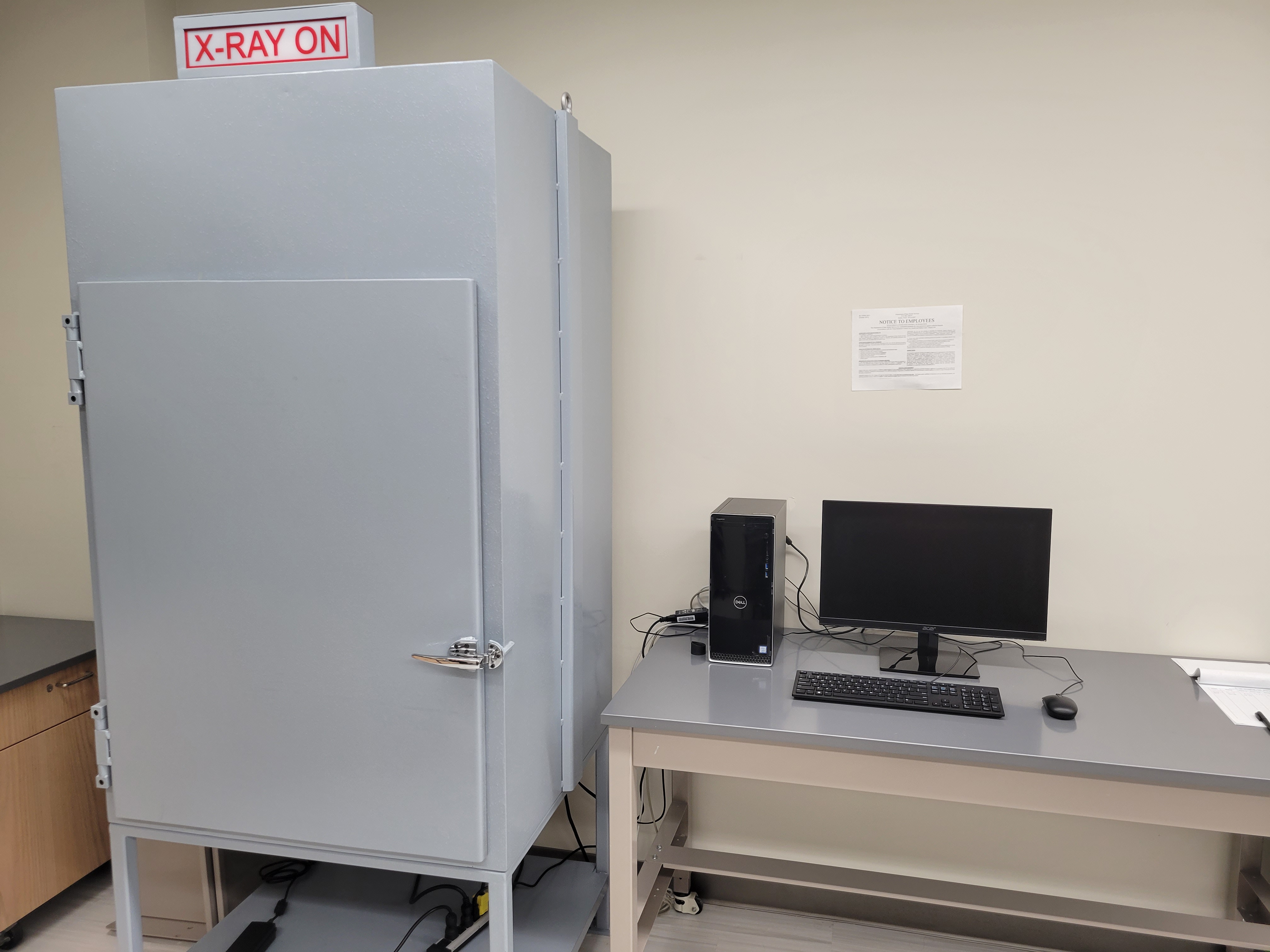
Digital X-Ray Machine
X-Ray Tube: Thermo Scientific PXSS-927 EA-R X-Ray Tube
Max Voltage: 90 kV
Max Power: 8 W
Spot Size: 5 microns
X-Ray Detector: Mars 1919V Flat Panel Detector
Resolution: 139 µm pixel pitch
The digital X-ray is ideal for studying tissues of preserved specimens. This non-destructive technique allows collection of vertebrate skeletal data without dissection. The detector is powerful, and with several working distances available within the cabinet, a wide range of specimen sizes can be used.
The cabinet, composed of lead compounds, reduces X-ray radiation to negligible amounts. Even still, students will need to take short radiation safety training to use the machine.
Cressington 108 Sputter Coater
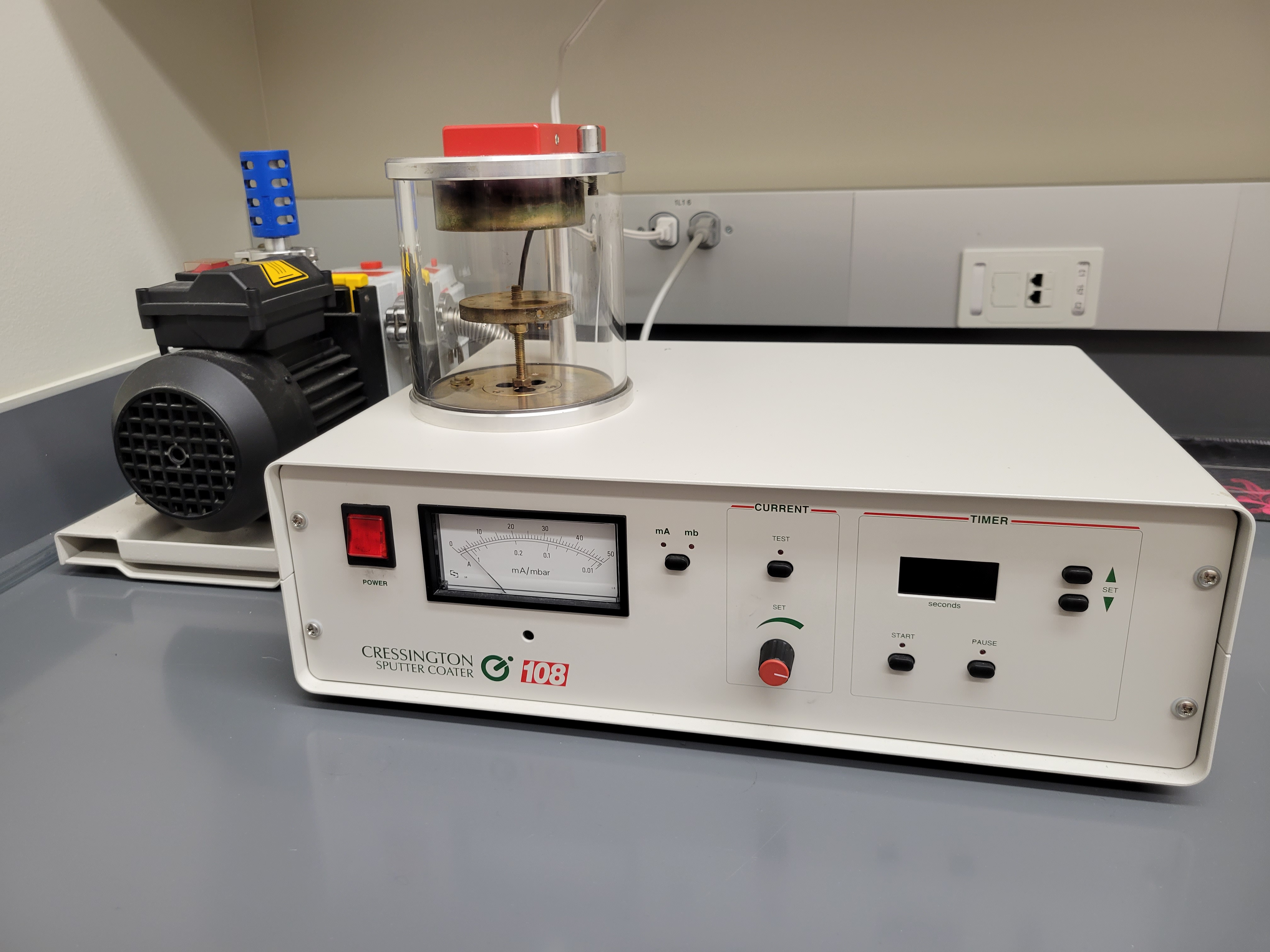
Electron microscopy can pose some complications for biological samples. Due to most organic material being made up of elements with a low atomic number, performing scanning electron microscopy on unprepared organic tissue could result in low signal and a bad image altogether. To compensate for this, sputter coaters are designed to deposit a fine film of gold particles on top of the sample studs. This thin layer of gold (sometimes platinum for higher magnification SEM) helps to distribute electron charge to improve contrast, as well as increase signal from secondary electrons.
The Cressington 108 Sputter Coater in the prep room is capable of coating 12 small sample studs in one cycle. Time spent sputtering, pressure, and sputter current can all be adjusted to accommodate a variety of sample surfaces.
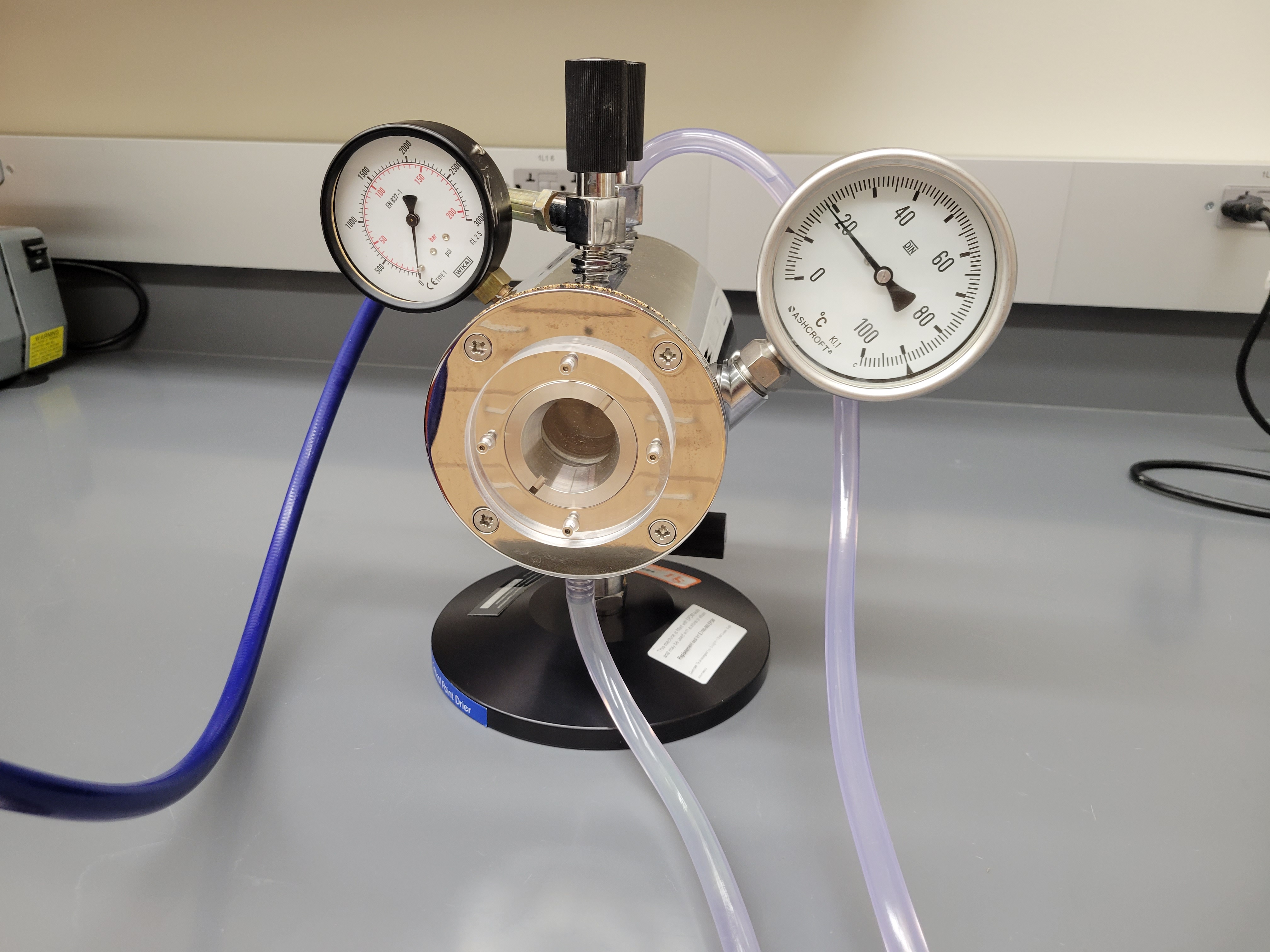
Quorum E3100 Critical Point Drier
Maximum # of sample baskets: 25 baskets
Lowest Temp. with ice bath: 9°C
Safety Burst: 1825 psi
For particularly small samples, a thin layer of gold won't be enough to produce good images with SEM. Many fine structures may experience warping when allowed to dry in the air, surface tension can cause tissues to shrivel up this way. Additionally, samples that are not completely dry may experience spot heating when exposed to the electron beam. Depending on the size of the sample and how dry it is, sometimes dramatic warping and sometimes burning of the sample can occur due to the intensity of the electron beam.
To account for this, the critical point drier is a completely manual apparatus that uses the critical point of carbon dioxide to exchange wet intermediate fluid in the samples for carbon. This enables small samples to completely dry without any artifacts during imaging from either air drying or high electron beam intensity.
Leica UC7 Ultramicrotome
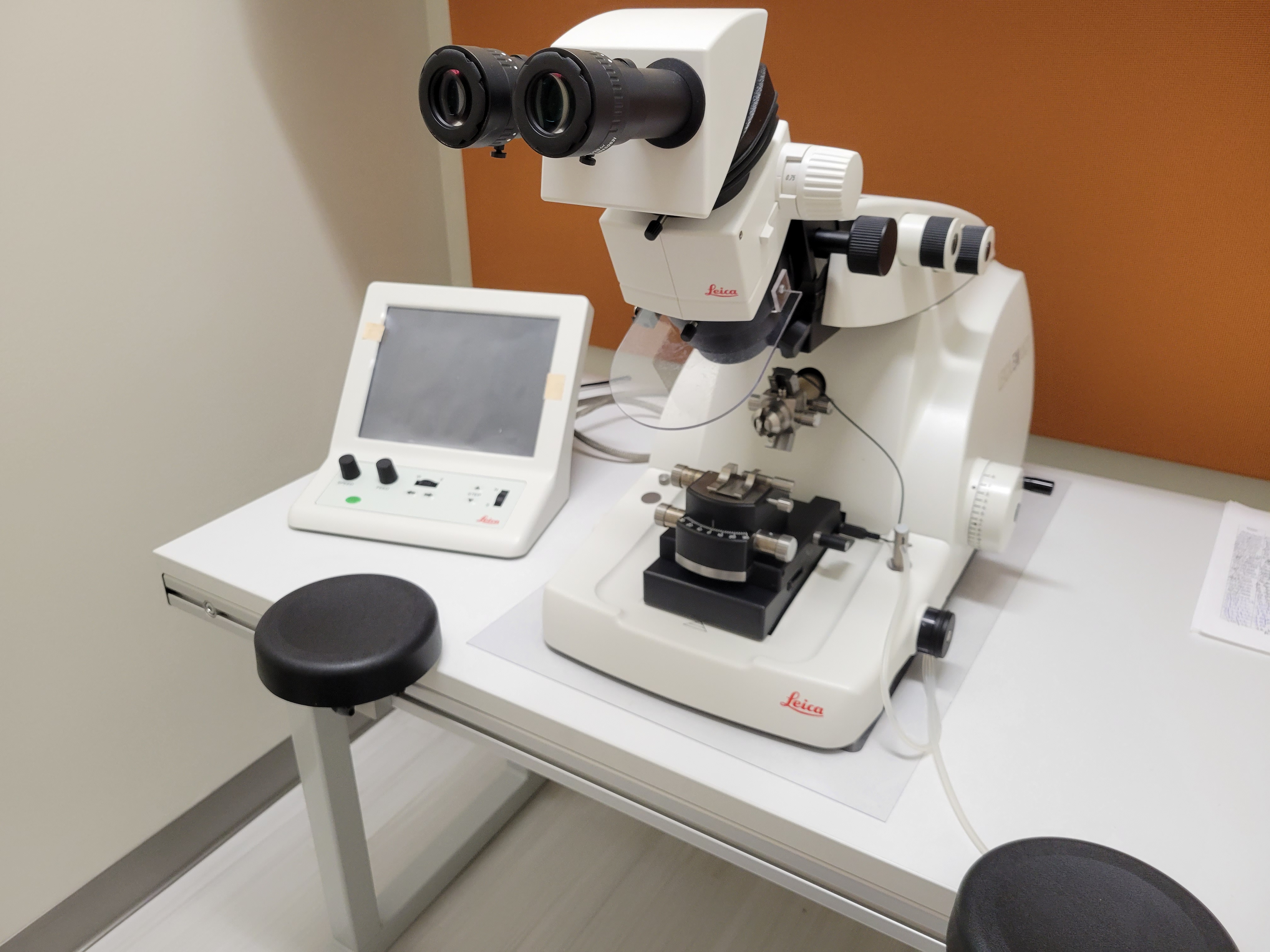
Section Range: 1µm - 50nm
Thin sectioning is necessary for both transmission electron microscopy and confocal microscopy techniques. Ultramicrotomy can achieve sections as thin as 50nm by slowly shaving off pieces of a resin block using a glass knife. These thin sections are much easier to work with than large samples which may be too thick for TEM, where the electron beam may not provide sufficient contrast. SEM and light microscopy can also benefit from sectioning. When studying a particular tissue or even organelle within a cell, it may be useful to cut several sections to assure that the best possible image is obtained.
Introduction to Microscopy
The majority of the teaching labs in the Life Sciences Building use basic transmitted light microscopes with the standard 4x, 10x, and 40x objective lenses. A number of histological stains are used to create contrast between different structures in a sample, whether that be individual cells or entire tissues. Our prep lab contains many of these stains for basic light microscopy.
If you encounter issues when using a microscope, please refer to some of the notes below. For the teaching scopes, lab assistants and graduate assistants should be handling any maintenance necessary. Students do not need to troubleshoot their own microscopes for risk of further damage to the objective lenses. Anything beyond the capability of lab assistants should be set aside for service. Notify Daniel Doucet (doucet@shsu.edu, LSB office 200K) for service on teaching scopes
Rates
|
Instruments |
SHSU Microscopy Center Higher Education Rate |
Average University Higher Education Rate |
Industry Rate |
|
Hitachi SU3500 Scanning Electron Microscope |
$30/hr |
$100+/hr |
$60/hr |
|
Hitachi H-7500 Transmission Electron Microscope |
$45/hr |
$100+/hr |
$80/hr |
|
Olympus Fluoview 3000 Confocal Microscope |
$20/hr |
$60+/hr |
$40/hr |
|
Digital X-ray |
No Charge |
$40/hr |
$40/hr |
|
Sputter coater |
$15/run |
$15/run |
$20/run |
|
Keyence VHX-5000 Digital Microscope |
No Charge |
$40/hr |
$20/hr |
|
Revolve 4 Echo microscope |
No Charge |
$40/hr |
$20/hr |
|
Training on SEM/TEM/confocal |
$50/hr (internal) / $180/hr (external) |
||
|
Sample preparation for SEM/TEM |
Contact us for pricing |
||
Contact Us
Visit Us
2000 Ave. I Rm 105
P.O. Box 2116
Huntsville, Texas 77341
Building Hours: 7:00 a.m. - 8:00 p.m., Monday - Friday
Call Us
936.294.1540
Email Us
Bio_Admin@shsu.edu